
Nazlee Sharmin, University of Alberta
Ava K. Chow, University of Alberta
Alice S. Dong, University of Alberta
Virtual microscopes are computer or web-based programs that enable users to visualize digital slides and mimic the experience of using a real light microscope. Traditional light microscopes have always been an essential teaching tool in health science education to observe and learn cell and tissue structures. However, studies comparing virtual and real light microscopes in education reported learners’ satisfaction with virtual microscopes regarding their usability, image quality, efficiency, and availability. Although the use of virtual or web-based microscopy is increasing, there is no equivalent decrease in the number of schools utilizing traditional microscopes. We conducted a scoping review to investigate the comparative impact of conventional and virtual microscopes on different aspects of learning. We report a relative effect of virtual and light microscopy on student performance, long-term knowledge retention, and satisfaction. Our results show that virtual microscopy is superior to traditional microscopes as a teaching tool in health science education. Further studies are needed on different learning components to guide the best use of virtual microscopy as a sole teaching tool for health care education.
Keywords: virtual microscope; web-based microscope; health science education; learning experience
Les microscopes virtuels sont des programmes informatiques ou web qui permettent aux utilisateurs de visualiser des diapositives numériques et d’imiter l’expérience de l’utilisation d’un vrai microscope optique. Les microscopes optiques traditionnels ont toujours été un outil d’enseignement essentiel dans l’enseignement des sciences de la santé pour observer et apprendre les structures cellulaires et tissulaires. Cependant, des études comparant les microscopes virtuels et optiques dans l’éducation ont rapporté la satisfaction des apprenants à l’égard des microscopes virtuels en ce qui concerne leur convivialité, leur qualité d’image, leur efficacité et leur disponibilité. Bien que l’utilisation de la microscopie virtuelle ou web augmente, il n’y a pas de diminution équivalente du nombre d’écoles utilisant des microscopes traditionnels. Nous avons effectué un examen de la portée pour étudier l’impact comparatif des microscopes conventionnels et virtuels sur différents aspects de l’apprentissage. Nous rapportons un effet relatif de la microscopie virtuelle et optique sur la performance des étudiants, la conservation des connaissances à long terme, et la satisfaction. Nos résultats montrent que la microscopie virtuelle est supérieure aux microscopes traditionnels en tant qu’outil d’enseignement dans le domaine de l’enseignement des sciences de la santé. D’autres études sont nécessaires sur différentes composantes d’apprentissage pour guider la meilleure utilisation de la microscopie virtuelle comme seul outil d’enseignement pour l’éducation en matière de soins de santé.
Mots-clés : microscope virtuel ; microscope basé sur le web ; enseignement des sciences de la santé ; expérience d’apprentissage
Traditional light microscopes have always been used for teaching tissue structures and microanatomy in health science education. Recent advancements in whole-slide imaging, virtual microscopes, digital slide viewers, and similar technologies show an immense possibility in education, with a potential to entirely substitute traditional light microscopes in many disciplines of health science education (Saco et al., 2016; Triola & Holloway, 2011). Virtual or web-based microscope refers to a computer or web-based program that enables users to mimic the experience of using a real light microscope. A virtual microscope allows users to view, navigate, and manipulate digital slides acquired from a camera-equipped microscope or a commercial digital slide scanning system (Triola & Holloway, 2011). The popularity and use of virtual microscopes are increasing throughout health professional education, especially in histology and pathology (Bloodgood & Ogilvie, 2006; Glatz-Krieger et al., 2006; Paulsen et al., 2010; Sharmin et al., 2021). Virtual microscopes improve the overall in-class teaching environment (Blake et al., 2003; Bloodgood & Ogilvie, 2006; Cotter, 2001; Kumar et al., 2004). Studies comparing virtual and real light microscopes in education found equal satisfaction from learners with the quality of image and usability while garnering greater satisfaction with the efficiency of learning and availability (Harris et al., 2001; Heidger et al., 2002; Kumar et al., 2004; Mills et al., 2007). Students’ academic performances are improved or unaffected by virtual microscopy (Harris et al., 2001; Helle et al., 2013; Kumar et al., 2004).
One of the critical advantages of a web-based or virtual microscope over a traditional light microscope is its ubiquitous availability (Triola & Holloway, 2011). This technology allows users to view digital slides anytime and at any place in the world, outside the classroom. The traditional method of teaching microanatomy and tissue structure relies on a limited number of light microscopes and glass slides, which does not allow simultaneous observation by multiple learners, prevents interactive in-class discussions. As per Capela et al. (2010), these limitations attenuate student motivation. Virtual microscopes enable multiple users to view the digital slides on a larger screen, promoting interactive discussion and team-based learning (Triola & Holloway, 2011). Digital slides and virtual microscopes can also be excellent resources for teaching. Instructors can pre-annotate slides outside class time and embed digital slides or links to specific views in teaching notes (Harris et al., 2011).
Although virtual microscopes and other computer-based slide-viewers provide access to many great-quality rare slides, they require computers, active Internet, or other smart devices, which may not be readily available to everyone. Although the use of virtual or web-based microscopy is increasing, there is not an equivalent decrease in the number of schools utilizing traditional microscopes for education (Bloodgood & Ogilvie, 2006), indicating that the questions regarding the impact of virtual microscopes in all aspects of learning are yet to be answered.
In this context, we aim to investigate the current scenario of virtual microscopy in health professional education, with a specific focus on the comparative impact of traditional and virtual microscopes in different aspects of learning. Our research question is: How do virtual microscopes compare with conventional microscopes in health science education?
Both medical and allied health databases were searched systematically to include all health professional education programs at the graduate and undergraduate levels. PubMed, The Cumulative Index to Nursing and Allied Health Literature (CINAHL) database, Excerpta Medica dataBASE (EMBASE), Cochrane, and Web of Science were searched systematically. Search terms were: virtual histology, virtual microscopy, and web-based microscopy combined with education, teaching, and learning. The details of search terms and search results are listed in Table 1.
Preferred Reporting Items for Systematic Reviews and Meta-Analyses (PRISMA) guidelines were followed to conduct the scoping review (Moher et al., 2009). Figure 1 represents the flow diagram of the study selection. Articles written in English and published in the last 10 years were included. To answer our research question, we included studies solely focused on health professional education. We excluded articles published before 2010, focused on non-health professional education, or applied virtual microscopy for diagnostic or clinical uses. We also excluded articles that are reviews, commentary, opinions, or technical descriptions. According to our established inclusion criteria, articles were screened based on titles and/or abstracts by two independent reviewers in the first round of review. In the second round, the two reviewers examined the full texts of the selected articles. After each review cycle, the disagreements between the first two reviewers were resolved by the third reviewer. Details of the inclusion and exclusion criteria are listed in Table 2.
Table 1
Detail of Search Terms and Search Results
| Search Term | Databases | ||||
| EMBASE | CINAHL | Cochrane | PubMed | Web of Science | |
| Web-based microscopy AND Education | 0 | 7 | 4 | 30 | 29 |
| Web-based microscopy AND Learning | 0 | 2 | 3 | 30 | 36 |
| Web-based microscopy AND Teaching | 0 | 1 | 2 | 38 | 21 |
| Virtual histology AND Education | 0 | 46 | 38 | 424 | 98 |
| Virtual histology AND Learning | 1 | 22 | 37 | 320 | 112 |
| Virtual histology AND Teaching | 0 | 25 | 31 | 388 | 110 |
| Virtual microscopy AND Learning | 2 | 7 | 23 | 107 | 188 |
| Virtual microscopy AND Education | 1 | 46 | 26 | 180 | 185 |
| Virtual microscopy AND Teaching | 0 | 11 | 26 | 338 | 167 |
| Total | 4 | 167 | 190 | 1,855 | 946 |
Table 2
Inclusion and Exclusion Criteria
| Selection Criteria | Inclusion | Exclusion |
| Language | English | Non-English |
| Year of study | Studies published within last 10 years | Studies published before 2010 |
| Study focus | Health professional education | Non- health professional education |
| Education: Didactic/theory/academic classroom teaching and learning | Clinical Use: Diagnostic/surgical/ clinical | |
| Undergraduate post-secondary education | K-12 education | |
| Examines user experience and/or learning outcomes | Descriptive and technical articles, review, commentary/opinion articles | |
| Study design | Any | Nil |
| Setting | Any | Nil |
The data from the 13 articles were extracted and organized (Table 3). The results relevant to the research question were then synthesized.
The initial search included 1,112 non-duplicate records from both medical and educational databases. The first screening phase by title and abstract retrieved 55 articles for full-text review. Forty-two reports were excluded in the second phase for not matching our research question or not conducting a comparative evaluation between virtual and conventional microscopes for education. Duplicates and conference proceedings were also excluded in this phase (Figure 1). After the two-phase screening, 13 articles were eligible for data extraction. All the qualified articles conducted a comparative assessment between traditional and virtual microscopes to teach health professional education.
All the studies included in our review divided research participants into either two or all of the following groups:
Groups using conventional microscopes
Groups using virtual microscopes
Groups using both traditional and virtual microscopes
Twelve studies evaluated students’ performance and knowledge attainment. Knowledge acquisition was assessed and compared between the groups from scores in formal board exams (Nauhria et al., 2019), online, multiple-choice, laboratory exams, identification tests (Lee et al., 2020), and score improvement from pre-test to post-test (Hande et al., 2017; Mione et al., 2013; Nauhria et al., 2019). Six studies assessed participants’ preferences and satisfaction on the Likert scale. Eleven studies took the quantitative, and two studies took the mixed-method approach to collect and analyze data from the participants.
Figure 1
Flow Diagram Explaining the Study Selection Process
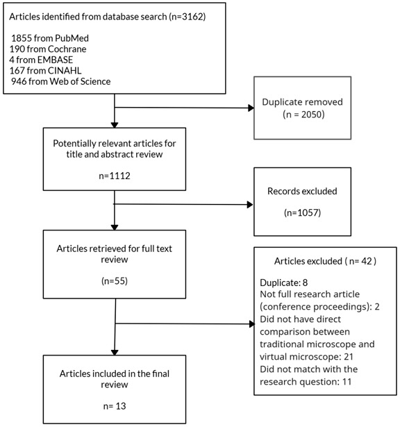
Table 3
Detail of the Studies Included in the Scoping Review
| Author, Year, Country | Research Method | Discipline of Health Science Education | Research Aim/Question | Research Participants | Brief Description of the Study | Key Findings | Theme |
| Mione et al., 2013 Belgium | Quantitative | Histology | To study the impact of the implementation of VM versus LM on the acquisition of histology knowledge. | The study included three different student populations: 1st-year bachelor students in Biomedical Sciences 2006-2007 (n = 172), 1st-year bachelor students in Biomedical Sciences 2007-2008 (n = 202), and 1st-year bachelor students in Logopaedic and Audiological Sciences 2007-2008 (n = 104). Total 478 | A pretest-posttest and cross-over design was adopted. In the first phase, students were divided into two groups. Group 1 performed the practical sessions with the LM. Group 2 performed the same sessions with the VM. In the second phase, the research subjects switched conditions. The prior knowledge levels of all students were assessed with a pre-test. Knowledge acquisition was measured with a post-test after each phase. | No significant differences were reported between pre-test and post-test scores of the student groups. Virtual microscopes are equivalent to optical (light) microscopes. | Knowledge acquisition |
| Lee et al., 2020 Taiwan | Mixed | Histology and Pathology | To examine the influence of VM on academic performance, and teacher and student perceptions. | 662 3rd-year medical and dental students studying histology and 651 4th-year students studying pathology | Students were divided into two groups. The light microscopy group used a LM in 2014 and 2015, while the light microscopy + virtual microscopy group used the VM platform and a LM in 2016 and 2017. Examination scores were compared. Participants were asked to complete a survey and write comments. | The light microscopy+ virtual microscopy group exhibited less score variability on laboratory examinations relative to their mean than the light microscopy group. Both teachers and students agreed that the virtual microscopy platform enhanced laboratory learning. | Academic performance |
| Nauhria et al., 2019 Grenada | Mixed | Pathology | To investigate whether VM or LM had a higher impact on student learning and performance in histopathology. Whether students preferred VM over LM. | 2nd-year medical students n = 152 | A sequential exploratory mixed method study design was used. A qualitative phase inquiring about student preference for VM or LM was followed by a randomized cross over study. Student preference was measured by an online survey based on a Likert scale. In the cross over study, students were randomized either to the VM or the LM arm, and their mean scores in standardized exams were compared after using VM and LM. | 83% of the students preferred to use VM over LM. Students who used VM scored significantly higher in both phases of the cross over study than those who used LM. | Academic performance Student satisfaction |
| Sagol et al., 2015 Turkey | Quantitative | Pathology | To evaluate the use of virtual microscopy in practical pathology sessions and its effects on students. | 2nd and 3rd-year medical students n = 351 | The practical sessions were carried out via virtual slides and the effect of the new technique was investigated at the end of each session. | The evaluation of the ratings showed that the students were easily adapted to the use of virtual microscopy. They found it user-friendly and thought that the opportunity of viewing slides at home was advantageous. | Student satisfaction |
| Ordi et al., 2015 Spain | Quantitative | Pathology | To determine the impact in student scores when moving from conventional microscopy to virtual microscopy. To assess the students’ impressions and changes in study habits regarding the impact of this tool. | Students from a medical school n = 181 | The authors evaluated two groups taking the discipline of pathology in the same course, one using conventional microscopy and the other virtual microscopy. The same set of slides used in the conventional microscopy classes was digitized and observed by the students using the Virtuoso viewer. The authors evaluated the skill level reached by the students with an online test. | There were no differences between the two groups in their marks in the online test. Students found the software friendly, easy-to-use, and effective. The most appreciated feature of virtual microscopy was the possibility to access images anywhere and at any time. | Knowledge acquisition Student satisfaction |
| Foad, 2016 Saudi Arabia | Quantitative | Pathology | To compare students’ apprehension of knowledge and skills via LM and VM. | 2nd-year medical students n = 40 | Students were randomly assigned to use either conventional, light, or virtual microscopy in practical sessions. The students’ apprehension of knowledge was assessed by written and practical exams. The authors also conducted pre- and post-test comparisons between VM and LM groups. | Students in the virtual microscopy group performed better than those in the light microscopy group as reflected by their more-uniform performance and less-scattered grades in both practical and written exams. Students from VM group showed significant improvement in their post-test scores compared to the other LM group. | Knowledge acquisition |
| Daniela et al., 2018 Chile | Quantitative | Histology | To evaluate the student’s academic performance and perception by learning muscle tissue module in the histology unit, using as a study tool light microscopy and a web application created for histological analysis. | 1st-year students of dentistry n = 92 | A total of 92 students were randomly divided into two groups. Group 1: 46 students used light microscopes. Group 2: 46 students used digital microscopy. At the end of the experimental phase, each group took a cognitive test which measured their ability to diagnose the various types of muscle tissue and to identify structures. A perception test was conducted after everyone had learned with both systems. | In the cognitive evaluation, the median grades were 5.4 for group 1 and 5.7 for group 2. In the perception survey, 73.24 % considered the VM evaluation fairer. It was concluded that the use of a VM tends to have better results than light microscopy. | Knowledge acquisition Student satisfaction |
| Brown et al., 2016 United Kingdom | Quantitative | Histology | To compare the effectiveness of virtual microscopy and traditional microscopy in teaching histology. | 3rd-year veterinary students from the two universities | 3rd-year veterinary students from two different schools completed a simple objective test, covering aspects of histology and histopathology, before and after a practical class covering relevant material presented as either glass slides viewed with a microscope or as digital slides. | There was an overall improvement in performance by students at both veterinary schools using both practical formats. Neither format was consistently better than the other, and neither school consistently outperformed the other. In comparing student appraisal of the use of digital slides and microscopes, digital technology was identified as having many advantages. | Knowledge acquisition Student satisfaction |
| Evans et al., 2020 USA | Quantitative | Cytology | To determine whether instruction using VM, compared to CM, is a successful method of training veterinary students to apply cytology in practice (i.e., sing light microscopes). | Veterinary students n = 71 | Students who attended a voluntary 3-hour cytology workshop were randomized to receive the same instruction with either VM (n = 35) or CM (n = 36). The control group (n = 22) of students who did not attend a workshop. All students took a post-workshop assessment involving the interpretation of four cases on glass slides with CM, designed to simulate cytology in general practice. | The mean assessment score of the VM group (14.18 points) was significantly higher than the control group, whereas the mean of the CM group was not significantly different from the controls. | Knowledge acquisition |
| Hande et al., 2017 India | Quantitative | Dental histology | To evaluate the effectiveness of virtual microscopy with conventional microscopy on student learning in dental histology. | Dental students n = 105 | Students were included and randomized into three groups: A, B, and C. Group A students studied the microscopic features of oral histologic lesions by conventional microscopy, Group B by virtual microscopy, and Group C by both traditional and virtual microscopy. The student’s understanding of the subject was evaluated by comparing pre- and post-test scores. | The difference in scores between Groups A, B, and C at pre-and post-test was highly significant. 87.61% of the students strongly agreed that the virtual microscopy was useful as a practically oriented teaching-learning tool and shows enhanced learning. | Knowledge acquisition Student satisfaction |
| Solberg, 2012 USA | Quantitative | Hematology | To examine student performance, skill retention and transferability, and self-efficacy beliefs amongst undergraduate MLS students learning cellular morphology with digital versus traditional slides. | Students from medical laboratory science (MLS) n = 74 | Participants were randomly assigned to either Group 1 or Group 2. Students in Group 1 used digital slides and in Group 2 used traditional slides for the myeloid maturation lab. Data were collected from three sources: immediate performance evaluation, a delayed performance evaluation, and a self-efficacy measure. | Students learning with digital slides performed better on assessments containing only traditional slide specimens than students learning with traditional slides, both immediately following the learning activity and after a considerable duration of time. Students learning with digital slides also reported slightly higher levels of self-efficacy related to cellular identification. | Knowledge acquisition Knowledge/ Skill retention Self-efficacy |
| Brueggeman et al., 2012 USA | Quantitative | Hematology | To evaluate the efficacy of virtual microscopy as the primary mode of laboratory instruction in undergraduate level clinical hematology teaching. | Students from medical laboratory science (MLS) n = 58 | Students were randomly assigned to either traditional microscopy or virtual microscopy instruction. Both groups had access to identical lecture materials. Students participated in three surveys requesting feedback on preparedness, perception, and expectations of the course before, during, and after delivery. All students took identical laboratory practical and written exams mid-term and at the end of the semester. | No significant differences between traditional microscopy and virtual microscopy groups with respect to group means for the midterm laboratory exam, the final laboratory exam, or the course total. | Academic performance |
| Tian et al., 2014 China | Quantitative | Histology | To describe a VM system for undergraduates and to evaluate the effects of promoting active learning and problem-solving skills. | 2nd-year medical students n = 221 | Students were divided into two groups. The VM group contained 115 students and was taught using the VM system. The LM group consisted of 114 students and was taught using the LM system. Post-teaching performances were assessed by multiple-choice questions, short essay questions, case analysis questions, and the identification of the structure of the tissue. Students’ teaching preferences and satisfaction were assessed using questionnaires. | Test scores in the VM group showed a significant improvement compared with those in the LM group. There were no substantial differences between the two groups in the mean score rate of multiple-choice questions and the short essay category; however, there were notable differences in the mean score rate of case analysis questions and identification of the tissue structure. The questionnaire results indicate that the VM system improves students’ productivity and promotes learning efficiency. | Academic performance Student productivity Learning efficacy |
Note. Virtual microscope = VM; Light microscope = LM; Conventional microscope = CM
The population included in the studies comprises students of medicine (5 studies), dentistry (2 studies), medicine and dentistry combined (1 study), biomedical sciences (1 study), veterinary (2 studies), and medical laboratory sciences (2 studies). The population is from the United States, United Kingdom, Belgium, Chile, China, Grenada, India, Saudi Arabia, Spain, Taiwan, and Turkey. An overview of the year the article was published and the demographic distribution of the population is shown in Figure 2. Virtual microscopy was applied to teach general histology (4 studies), pathology (4 studies), both histology and pathology (1 study), cytology (1 study), dental histology (1 study), and hematology (2 studies).
Figure 2
Distribution of Articles Based on Year of Publication and Location of the Population

Assessing the differences between pre-test and post-test scores is frequently used to evaluate students’ performance. Pre-test-post-test design involves collecting pre-test measures of the outcome of interest before administering some treatment, followed by a post-test on the same measure after treatment or intervention occurs. Pre-test-post-test designs can be applied in experimental and quasi-experimental educational research and used with or without control groups (Salkind, 2010). Mione et al. (2013) divided students into the light microscope and virtual microscope groups. The authors compared the differences in pre- and post-test scores between the groups. No significant differences were reported between pre-test and post-test scores among the student groups (2013).
On the other hand, Hande et al. (2017) divided students into three groups who were learning either with conventional microscopes (Group A), virtual microscopes (Group B), or both (Group C). Comparing the difference between pre-test and post-test scores among groups showed significant differences. Handle et al. discovered that the students who used both the light and virtual microscopes demonstrated the most significant improvement in their post-test scores compared to the pre-test, followed by the groups who used only virtual microscopes as their learning tool. The student group who used only conventional light microscopes showed the slightest difference between their pre- and post-test scores (2017).
Student performances in the formal academic examinations were also recorded to compare groups studying with conventional or virtual microscopes. Lee et al. (2020) divided students into two groups where one group used only light microscopes, and the other group used both light and virtual microscopes for learning histology and pathology. The group of students who used light and virtual microscopes performed better with less score variability on their laboratory examinations than the group who used only light microscopes. In a cross over study, Nauhria et al. (2019) randomized participants either to the virtual microscopy or the light microscopy group then compared their mean scores in standardized exams. Students who used virtual microscopy scored significantly higher in the national board exam than those who used light microscopy. The authors also conducted pre- and post-test comparisons, where the students from the virtual microscopy group showed significant improvement in their post-test scores compared to the light microscopy group.
Ordi et al. (2015) used an online test to comparatively evaluate the skill acquired by two student groups. One group used light microscopes, and the other used virtual microscopes for learning pathology. The study found no significant differences in students’ performance between the groups. Similarly, no significant differences were reported by Brueggeman et al. (2012) concerning group mean scores for the midterm laboratory exam, final laboratory exam, or the course total. However, in the study by Foad (2016), students from the virtual microscopy group performed better than those in the light microscopy group in practical and written exams. Compared to the conventional microscope group, students using virtual microscopes scored higher in multiple-choice questions, short essays, case analyses, and cognitive tests, which measured their abilities to diagnose tissue structures from histological slides (Daniela et al., 2018; Tian et al., 2014).
In summary, 10 out of 12 studies found that students learning with virtual microscopes performed better in knowledge acquisition tests, including post-test, national board exam, online, written, multiple-choice, case analysis, and structure identification. The other two studies reported no significant differences in the mean score between groups using virtual or light microscopes (Table 3).
In a non-experimental comparative study, Solberg (2012) randomly assigned students in a clinical hematology course to use digital slides in virtual microscopes (Group 1) and traditional glass slides in the light microscope (Group 2). To evaluate students’ immediate performance, separate microscope stations were set up where the students were asked to identify cellular structures. A delayed performance evaluation was conducted nine weeks after the initial laboratory sessions, similar to the immediate assessment. Students learning with the virtual microscope and digital slides (Group 1) performed better on evaluations immediately following the learning activity and after a considerable time (2012).
Surveys based on the Likert scale were used to assess student preferences between virtual and conventional microscopes and their overall satisfaction with virtual microscopy and digital slides. In the study by Nauhria et al. (2019), 83% of the students preferred to use a virtual microscope over a light microscope. Student participants showed a high level of satisfaction and found virtual microscopes user-friendly and effective as teaching and learning tools (Daniela et al., 2018; Hande et al., 2017; Ordi et al., 2015; Sagol et al., 2015; Tian et al., 2014). The ability to view slides anywhere and at any time was considered highly advantageous by the participants (Brown et al., 2016; Ordi et al., 2015; Sagol et al., 2015).
The goal of our scoping review was to investigate and synthesize a comparative overview of the impact of virtual and light microscopes as a teaching and learning tool for health science education. The eligible studies for our scoping review primarily evaluated student performance in knowledge tests and their satisfaction and preferences. We did not assess the quality of the studies included in our scoping review (Arksey et al., 2005). There is a clear need to conduct further studies to investigate the impact of virtual microscopy on other learning components, including engagement, knowledge retrieval, self-regulated learning, self-efficacy, and motivation. The findings of our review was based on the small number of articles that met our inclusion criteria specifically relevant to our research question. We acknowledge that negative results may have been missed due to publication bias.
The use and popularity of virtual microscopes appear to be increasing worldwide across different disciplines of health science education. The vast majority of published literature on virtual microscopes focuses on the technical details of the technology and its sole effect on education. Our scoping review aimed to generate an overview comparing the impact of virtual and light microscopes in health professional education. We included comparative evaluation studies between student groups using conventional and virtual microscopes.
Based on our data extraction, we have summarized the effect of virtual microscopy on student performance, long-term knowledge retention, and satisfaction. The literature included comparative evaluations between groups of learners who used (i) only conventional microscopes, (ii) only virtual microscopes, and (iii) a combination of conventional microscopes. All the articles that assessed student satisfaction with virtual micoscopes reported it at a high level. Ten out of twelve studies reported higher test scores and better performance in knowledge acquisition by the student groups who used virtual microscopes alone or in combination with light microscopes. Two studies reported no significant differences between the light and virtual microscope users regarding group means for the midterm laboratory exam (Brueggeman et al., 2012) and online test (Ordi et al., 2015). These findings indicate that virtual microscopes have either equally good or better effects on students’ knowledge acquisition or academic performance compared to light microscopes. In addition to the panning and zooming features, virtual microscopes also offer the ability to highlight areas of interest and create thumbnail views or location boxes for tracking navigation, which positively affects students learning. This technology can also potentially increase students’ basic knowledge and problem-solving skills (Tian et al., 2014).
Only one study in our review collected test scores from participants who used virtual microscopes alone or virtual microscopes together with light microscopes (Hande et al., 2017). The study found that students who used both light and virtual microscopes performed significantly better than the group who used only a virtual microscope or only a light microscope as a learning tool. This performance indicates that virtual microscopes can augment traditional light microscopes in enhancing the learning experience and grasp of the subject (2017).
Only 13 studies fulfilled our selection criteria, suggesting that although there is widespread adoption of virtual microscopes as a teaching modality, there are not enough studies exploring the impact of this technology compared to light microscopes. The potential and benefits of virtual microscopes appear evident. However, further studies in this area can guide the best use of this technology to support and enhance active learning, engagement, professional development, problem-solving skills, and satisfaction.
Virtual microscopes are gaining popularity as a teaching tool for microanatomy and pathology. Reportedly, educational institutions are replacing traditional light microscopes with virtual systems to cope with increasing costs, space, and equipment. We conducted a scoping review to identify the relative effect of virtual and light microscopy on student performance, long-term knowledge retention, and satisfaction. All studies included in our review reported virtual microscopy as superior or equal to light microscopes in all aspects of students’ learning experiences. Further studies in this area can guide educational institutions and educators to identify the best use of virtual microscopes as a teaching tool.
Arksey, H., & O’Malley, L. (2005). Scoping Studies: Towards a methodological framework. International Journal of Social Research Methodology: Theory & Practice, 8(1), 19-32. https://doi.org/10.1080/1364557032000119616
Blake, C. A., Lavoie, H. A., & Millette, C. F. (2003). Teaching medical histology at the University of South Carolina School of Medicine: Transition to virtual slides and virtual microscopes. Anatomical Record. Part B, New Anatomist, 275(1), 196-206. https://doi.org/10.1002/ar.b.10037
Bloodgood, R. A., & Ogilvie, R. W. (2006). Trends in histology laboratory teaching in United States medical schools. Anatomical Record. Part B, New Anatomist, 289(5), 169-175. https://doi.org/10.1002/ar.b.20111
Brown, P. J., Fews, D., & Bell, N. J. (2016). Teaching veterinary histopathology: A comparison of microscopy and digital slides. Journal of Veterinary Medical Education, 43(1), 13-20. https://doi.org/10.3138/jvme.0315-035R1
Brueggeman, M. S., Swinehart, C., Yue, M. J., Conway-Klaassen, J. M., & Wiesner, S. M. (2012). Implementing virtual microscopy improves outcomes in a hematology morphology course. American Society for Clinical Laboratory Science, 25(3), 149-155. https://doi.org/10.29074/ascls.25.3.149
Capela e Silva, F., Rato, L. M., & Lopes, O. S. (2010). Learning and teaching histology: Traditional and computational methods. Proceedings of International Conference “Learning and Teaching in Higher Education”; Apr 15-16; Evora, Portugal. http://hdl.handle.net/10174/2094
Cotter J. R. (2001). Laboratory instruction in histology at the University at Buffalo: Recent replacement of microscope exercises with computer applications. The Anatomical Record, 265(5), 212-221. https://doi.org/10.1002/ar.10010
Daniela, B., Melisa, G., Luis, A. J., Cristian, A., Jorge, T., Rosario, M., & Natividad, S. (2018). Academic achievement and perception of two teaching methods in histology: Light and digital microscopy. Pilot study. International Journal of Morphology, 36(3).
Evans, S. J., Moore, A. R., Olver, C. S., Avery, P. R., & West, A. B. (2020). Virtual microscopy is more effective than conventional microscopy for teaching cytology to veterinary students: A randomized controlled trial. Journal of Veterinary Medical Education, 47(4), 475-481. https://doi.org/10.3138/jvme.0318-029r1
Foad A. (2016). Comparing the use of virtual and conventional light microscopy in practical sessions: Virtual reality in Tabuk University. Journal of Taibah University Medical Sciences, 12(2), 183-186. https://doi.org/10.1016/j.jtumed.2016.10.015
Glatz-Krieger, K., Spornitz, U., Spatz, A., Mihatsch, M. J., & Glatz, D. (2006). Factors to keep in mind when introducing virtual microscopy. Virchows Arch. 448: 248-255. https://doi.org/10.1007/s00428-005-0112-2
Hande, A. H., Lohe, V. K., Chaudhary, M. S., Gawande, M. N., Patil, S. K., & Zade, P. R. (2017). Impact of virtual microscopy with conventional microscopy on student learning in dental histology. Dental Research Journal, 14(2), 111. https://pubmed.ncbi.nlm.nih.gov/28584534
Harris, T., Leaven, T., Heidger, P., Kreiter, C., Duncan, J., & Dick, F. (2001). Comparison of a virtual microscope laboratory to a regular microscope laboratory for teaching histology. The Anatomical Record, 265(1), 10-14. https://doi.org/10.1002/ar.1036
Heidger, P. M., Jr, Dee, F., Consoer, D., Leaven, T., Duncan, J., & Kreiter, C. (2002). Integrated approach to teaching and testing in histology with real and virtual imaging. The Anatomical Record, 269(2), 107-112. https://doi.org/10.1002/ar.10078
Helle, L., Nivala, M., & Kronqvist, P. (2013). More technology, better learning resources, better learning? Lessons from adopting virtual microscopy in undergraduate medical education. Anatomical Sciences Education, 6(2), 73-80. https://doi.org/10.1002/ase.1302
Kumar, R. K., Velan, G. M., Korell, S. O., Kandara, M., Dee, F. R., & Wakefield, D. (2004). Virtual microscopy for learning and assessment in pathology. The Journal of Pathology, 204(5), 613-618. https://doi.org/10.1002/path.1658
Lee, B. C., Hsieh, S. T., Chang, Y. L., Tseng, F. Y., Lin, Y. J., Chen, Y. L., Wang, S. H., Chang, Y. F., Ho, Y. L., Ni, Y. H., & Chang, S. C. (2020). A web-based virtual microscopy platform for improving academic performance in histology and pathology laboratory courses: A pilot study. Anatomical Sciences Education, 13(6), 743-758. https://doi.org/10.1002/ase.1940
Mills, P. C., Bradley, A. P., Woodall, P. F., & Wildermoth, M. (2007). Teaching histology to first-year veterinary science students using virtual microscopy and traditional microscopy: A comparison of student responses. Journal of Veterinary Medical Education, 34(2), 177-182. https://doi.org/10.3138/jvme.34.2.177
Mione, S., Valcke, M., & Cornelissen, M. (2013). Evaluation of virtual microscopy in medical histology teaching. Anatomical Sciences Education, 6(5), 307-315. https://doi.org/10.1002/ase.1353
Moher, D., Liberati, A., Tetzlaff, J., Altman, D. G., & PRISMA Group (2009). Preferred reporting items for systematic reviews and meta-analyses: The PRISMA statement. PLoS Medicine, 6(7), e1000097. https://doi.org/10.1371/journal.pmed.1000097
Nauhria, S., & Ramdass, P. (2019). Randomized cross-over study and a qualitative analysis comparing virtual microscopy and light microscopy for learning undergraduate histopathology. Indian Journal of Pathology & Microbiology, 62(1), 84-90. https://doi.org/10.4103/IJPM.IJPM_241_18
Ordi, O., Bombí. J. A., Martínez, A., Ramírez, J., Alòs, L., Saco, A., Ribalta, T., Fernández, P. L., Campo, E., & Ordi, J. (2015). Virtual microscopy in the undergraduate teaching of pathology. J Pathol Inform, 6(1). https://doi.org/10.4103/2153-3539.150246
Paulsen, F. P., Eichhorn, M., & Bräuer, L. (2010). Virtual microscopy: The future of teaching histology in the medical curriculum? Annals of Anatomy = Anatomischer Anzeiger : Official Organ of the Anatomische Gesellschaft, 192(6), 378-382. https://doi.org/10.1016/j.aanat.2010.09.008
Saco, A., Bombi, J. A., Garcia, A., Ramírez, J., & Ordi, J. (2016). Current status of whole-slide imaging in education. Pathobiology: Journal of Immunopathology, Molecular and Cellular Biology, 83(2-3), 79-88. https://doi.org/10.1159/000442391
Sağol, Ö., Yörükoğlu, K., Lebe, B., Durak, M. G., Ulukuş, Ç., Tuna, B., Musal, B., Canda, T., & Özer, E. (2015). Transition to virtual microscopy in medical undergraduate pathology education: First experience of Turkey in Dokuz Eylül University Hospital. Turk Patoloji Dergisi, 31(3), 175-180. https://doi.org/10.5146/tjpath.2015.01329
Salkind, N. J. (2010). Encyclopedia of research design (Vols. 1-0). Thousand Oaks, CA: SAGE Publications, Inc. http://doi: 10.4135/9781412961288
Sharmin, N., Chow, A. K., Dong, A. S., & Milos, N. C. (2021). Histoscope: A web-based microscopy tool for oral histology education. Healthcare Informatics Research, 27(2), 146-152. https://doi.org/10.4258/hir.2021.27.2.146
Solberg, B. L. (2012). Digital and traditional slides for teaching cellular morphology: A comparative analysis of learning outcomes. Clinical Laboratory Science, 25(4), 4-12.
Tian, Y., Xiao, W., Li, C., Liu, Y., Qin, M., Wu, Y., Xiao, L., & Li, H. (2014). Virtual microscopy system at Chinese medical university: An assisted teaching platform for promoting active learning and problem-solving skills. BMC Medical Education, 14(1), 1-8. https://doi.org/10.1186/1472-6920-14-74
Triola, M. M., & Holloway, W. J. (2011). Enhanced virtual microscopy for collaborative education. BMC Medical Education, 11(4). https://doi.org/10.1186/1472-6920-11-4
Nazlee Sharmin is an Assistant Teaching Professor at the School of Dentistry at the University of Alberta in Canada. She completed her PhD in Physiology, Cell, and Developmental Biology and a MEd from the University of Alberta. Her research interest focuses on developing technologies in classroom teaching to improve students’ learning experiences. Email: nazlee@ualberta.ca ORCID: https://orcid.org/0000-0002-2408-2333
Ava K. Chow is an Associate Professor at the School of Dentistry at the University of Alberta in Canada. She completed her PhD in Medical Sciences and a MEd from the University of Alberta. Her research interests include examining technology in education, foundational science education, and student-wellness. In lab, she studies the linkages between oral and systemic health. Email: akchow@ualberta.ca ORCID: https://orcid.org/0000-0002-1972-0310
Alice S. Dong has completed her Doctor of Dental Surgery from the School of Dentistry, University of Alberta in Canada. She is currently practicing as a general dentist. Email: sd5@ualberta.ca ORCID: https://orcid.org/0000-0002-2620-033X